graph TD

classDef start fill:#1b5e20,color:#ffffff,stroke:#66bb6a;
classDef step fill:#0d47a1,color:#ffffff,stroke:#42a5f5;
classDef decision fill:#4a148c,color:#ffffff,stroke:#ab47bc;
classDef alert fill:#b71c1c,color:#ffffff,stroke:#ef5350;

A(<b>Cold Exposure</b><br>Initial Assessment):::start
B{<b>Injury Category</b>}:::decision

A --> B

B -->|Systemic| C[<b>Accidental Hypothermia</b><br>Core temp under 35C]:::step
B -->|Localized| D[<b>Soft Tissue Injury</b><br>Extremities and exposed skin]:::step
B -->|Recurrent| E[<b>Genetic Syndromes</b><br>Investigate for auto-inflammation]:::step

C --> F{<b>Clinical Stage</b>}:::decision

F -->|Mild 32 to 35C| G[<b>Mild Hypothermia</b><br>Shivering intact]:::alert
F -->|Mod 28 to 32C| H[<b>Moderate Hypothermia</b><br>Stupor and decreased pulse]:::alert
F -->|Severe Under 28C| I[<b>Severe Hypothermia</b><br>Coma and bradycardia]:::alert

G --> M[<b>Passive Rewarming</b><br>Remove wet clothes and insulate]:::step
H --> N[<b>Active Rewarming</b><br>Forced air and warmed IV fluids]:::step
I --> O[<b>Internal Rewarming</b><br>Check pulse 1 min and warm blood]:::step

D --> J{<b>Tissue Status</b>}:::decision

J -->|Freezing| K[<b>Frostbite I to IV</b><br>Ice crystal formation]:::alert
J -->|Non Freezing| L[<b>Frostnip or Chilblains</b><br>Edema and no necrosis]:::alert

K --> P[<b>Rapid Rewarming</b><br>Circulating bath 37 to 39C]:::step
L --> Q[<b>Conservative Mgmt</b><br>Rewarm and topical creams]:::step
General Principles
Occurs when physiological heat generation fails to overcome environmental heat loss.
Develops even at temperatures above 0°C.
Heat transfers to environment via radiation, conduction, convection, respiration, and evaporation.
Categorized broadly into systemic injuries (accidental hypothermia) and localized soft tissue injuries (freezing and non-freezing).
Systemic Injury: Accidental Hypothermia
Definition And Pathophysiology
Defined as unintentional core body temperature drop below 35°C.
Cerebral function diminishes at 33-34°C.
Early manifestations include irritability, confusion, and poor decision-making.
Progresses to lethargy, somnolence, and coma.
Clinical Staging And Characteristics
State
Core Temperature
Clinical Characteristics
Mild
32-35°C
Increased shivering thermogenesis; increased metabolic rate; amnesia and dysarthria; ataxia; apathy; normal blood pressure.
Moderate
28-32°C
Stupor; 25% decrease in oxygen consumption; decreased shivering thermogenesis; atrial fibrillation and other dysrhythmias; pulse and cardiac output reduced to two-thirds normal.
Severe
<28°C
Coma with loss of cerebrovascular autoregulation; severe bradycardia; hypotension; high risk of unstable tachycardias (ventricular fibrillation) and asystole.
Emergency Management
General Measures
Handle gently and keep horizontal to prevent cardiovascular collapse.
Remove wet clothing immediately.
Replace with dry clothing and insulation to halt heat loss.
Mild Hypothermia Management (32-35°C)
Initiate passive rewarming using insulation and vapor barriers.
Provide active external rewarming with heat packs applied to neck, chest, upper torso, axilla, and groin.
Protect exposed skin from direct burns.
Support shivering with high-calorie oral fluids and carbohydrates if child remains alert.
Moderate Hypothermia Management (28-32°C)
Deploy active external rewarming to upper torso, chest, axilla, and back utilizing forced-air systems or large heat pads.
Administer intravenous or intraosseous fluids containing glucose, warmed to 40-42°C.
Maintain continuous cardiac monitoring due to high risk of unstable arrhythmias from cold heart and acidosis.
Transfer hemodynamically unstable patients to facilities capable of extracorporeal membrane oxygenation.
Severe Hypothermia (<28°C) And Cardiac Arrest
Assess pulse for up to 1 minute before initiating cardiopulmonary resuscitation.
Utilize bedside echocardiogram to detect organized electrical activity.
Initiate cardiopulmonary resuscitation for absent cardiac activity.
Never withhold cardiopulmonary resuscitation based on temperature unless fatal injury exists or chest compression proves impossible.
Hold vasoactive medications until core temperature exceeds 30°C.
Administer vasoactive medications at twice normal dosing interval between 30°C and 35°C.
Attempt single defibrillation or cardioversion at maximum power.
Hold further electrical shocks until core temperature exceeds 30°C.
Initiate active internal rewarming via warm intravenous fluids, extracorporeal blood warming, or hemodialysis alongside extracorporeal membrane oxygenation.
Localized Soft Tissue Cold Injuries
Freezing Cold Injury: Frostbite
Pathogenesis
Occurs at or below freezing temperatures.
Progresses through four phases: prefreeze, freeze-thaw, vascular stasis, and late ischemic.
Cellular destruction results from intracellular and extracellular ice crystal formation during freeze-thaw phase.
Exacerbated by ischemic-reperfusion injury and microvascular thrombosis.
Clinical Grading Of Frostbite
Grade
Field Classification
Clinical Features
Grade I
Superficial
Superficial injury; edema and redness without necrosis; numbness; firm white-yellow plaque; no blisters.
Grade II
Superficial
Substantial edema and erythema; clear or milky fluid-filled vesicles and blisters; desquamation forms black eschar.
Grade III
Deep
Extends into dermis and vascular plexus; hemorrhagic deeper blisters; blue-gray discoloration; skin necrosis.
Grade IV
Deep
Full-thickness freezing of skin, subcutaneous tissue, muscle, tendon, and bone; little edema; mottled red progressing to dry, black, mummified tissue; requires amputation.
Management Of Frostbite
Protect injured area from cold exposure.
Remove constricting items including jewelry.
Strictly prevent refreezing if spontaneous thawing commences.
Initiate rapid rewarming using circulating water bath heated to 37-39°C for 30 minutes.
Leave clear blisters intact.
Never aspirate hemorrhagic bullae.
Administer pain control medications.
Ensure updated tetanus prophylaxis.
Consider adjuvant therapies for severe cases 12-72 hours post-thawing.
Adjuvant options include vasodilators, antiplatelet drugs, synthetic prostacyclin analogues, or intra-arterial tissue plasminogen activator.
Non-Freezing Cold Injuries
Frostnip
Associated with vasoconstriction and superficial ice crystal formation.
Presents with localized numbness and pallor.
Lacks cellular damage.
Resolves rapidly upon external warming.
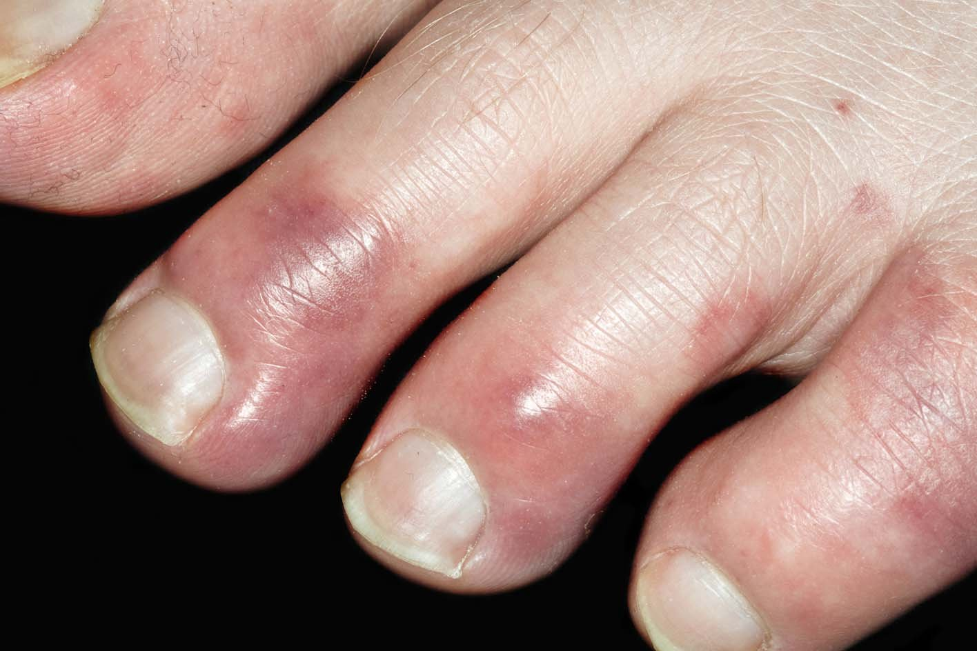
Chilblains (Pernio)
Idiopathic condition triggered by cold, damp exposure.
Presents as painful, edematous, bluish-red papular or nodular lesions.
Affects acral locations including fingers, toes, and ears.